FRC Conduit Carries Chemokines and Soluble Antigens to DC & HEV in Lymph Node Cortex

Larger animated FRCC image
See Intravital Multispectral 2-photon images of Lymphocytes Moving on Fibroblastic Reticular Cell Surfaces in the Lymph Node Cortex
Back to Previous Page With Lymphocyte Emigration Across HEV Walls
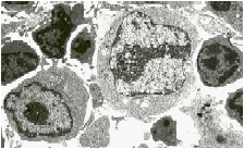
T-cell size correlates with activation state. Small are resting, Large are very active and may divide, and Intermediate are highly motile transitional forms

TEM of lymphocyte morphologies at stages from rest (sm) to activation (l).

Studies of what directs T- and B-cells after they get across HEV walls were guided by a visual finding. The main connective tissue structures that form a scaffold for lymphocytes to crawl around the cortex also appear to manufacture and conduct cytokines and other soluble materials in a fiber-filled tube that we call the "fibroblastic reticular cell conduit (FRCC)." Flow in the conduit has directionality and moves from lymph sinuses toward the walls of venules where lymphocytes emigrate. Thus FRCCs deliver the message in addition to providing the highway for migration of cells leaving the blood. Cells which do not encounter antigens or other stimuli that might cause them to change behavior and divide, leave by crawling into efferent lymphatic channels where they are returned to the bloodstream to recirculate again. * The FRC Conduit is also involved in viral pathogenesis Davis et al,1997. See also: Tara Waterman's WebSite on Ebola; David Sander's Filoviridae and Images Pages; our articles on the FRC Conduit in Viral Hemorrhagic Diseases and CD45 reduction making mice resistant to ebola.

FRC Conduit References

(Click HERE for our J Exp Med article on FRCC & Chemokines

The animated drawing (above) represents uptake and distribution of chemokines in FRCC after they arrive via afferent lymph. The drawing was constructed after studying sequential distribution of horseradish peroxidase tracer over a period of time less than 3 minutes. See Amer. J. Path. 80:387-418, 1975.

References

1. Anderson, A. O., N. D. Anderson. 1975. Studies on the structure and permeability of the microvasculature in normal rat lymph nodes. Amer. J. Path. 80:387-418.

2. Anderson, A. O. and N. D. Anderson. 1976. Lymphocyte emigration from high endothelial venules in rat lymph nodes. Immunology 31:731-748.

3. Ebnet, K., E.P. Kaldjian, A.O. Anderson, and S. Shaw, 1996, Orchestrated Information Transfer Underlying Leukocyte Endothelial Interactions. Annu. Rev. Immunol. 14:155-177.

4. Gretz, J.E., E.P. Kaldjian, A.O. Anderson, and S. Shaw, 1996,Sophisticated strategies for information encounter in the lymph node: the reticular network as a conduit of soluble information and a highway for cell traffic. J.Immunol. 157:495-499.

5. Gretz, J.E., A.O. Anderson, and S. Shaw, 1997 "Cords, Channels, Corridors, and Conduits: Critical architectural elements facilitating cell interactions in the lymph node cortex." Immunol. Rev. 156:11-24

Home | Recirculation | Emigration at HEV | Locomotion | Chemotaxis & Links

Dendritic Cells in TDL | Dendritic cells in Tissue | Immunology Lecture Notes

See who's visiting this page.View Page Stats